QUICK ANSWER Botox is a brand of botulinum toxin type A, a prescription medicine that temporarily reduces the activity of selected...
What That Eye Scan Actually Shows: A Guide to Eye Diagnostic Tests
By Dr. Joe Kwarteng
Senior Ophthalmologist, St. Thomas Eye Hospital

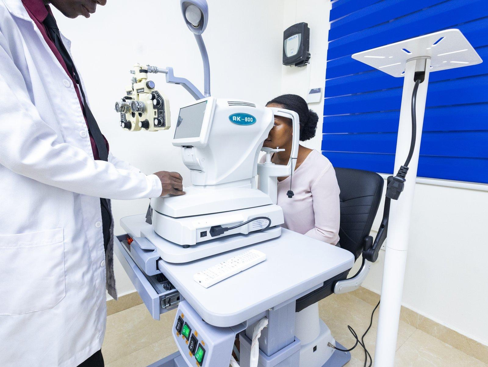
You sit down. A machine hums. A light flashes. The ophthalmologist looks at a screen you can’t quite see and says something like “your RNFL looks fine” or “we need a macular OCT.”
Most patients nod along and never ask what actually just happened to their eye.
That’s a problem because diagnostic imaging is where modern ophthalmology earns its keep. A slit lamp and a vision chart can tell you a lot, but they can’t see inside the eye in the way that matters for catching glaucoma, diabetic retinopathy, or a retinal tear before they cause permanent damage.
This guide explains, in plain language, what each diagnostic test at St. Thomas Eye Hospital actually does, what it can detect, and why your ophthalmologist might order one over another.
OCT: Optical Coherence Tomography
What it does: Takes a cross-sectional, almost 3D image of your retina; think of it as an ultrasound that uses light instead of sound waves. It’s painless, takes seconds, and requires no injections.
What it catches: OCT is the single most important tool for early glaucoma detection. It measures the thickness of the retinal nerve fibre layer (RNFL) — the layer of cells that carries signals from your eye to your brain. In glaucoma, this layer thins years before you notice any change in your visual field. OCT can flag that thinning while your vision still feels completely normal.
We offer two versions of this test:
• Glaucoma OCT (RNFL analysis): Measures the nerve fibre layer around the optic nerve.
• Combined Glaucoma OCT (RNFL + GCA): Adds ganglion cell analysis, looking at a deeper layer of cells that can show damage even earlier than the RNFL alone.
• Macular OCT: Focused on the centre of the retina, used to detect diabetic macular oedema, age-related changes, and other conditions affecting central vision.
If you have diabetes or a family history of glaucoma, expect OCT to be part of your routine screening. We explain why this matters so much for diabetic patients in our diabetic retinopathy guide.
FFA: Fundus Fluorescein Angiogram
What it does: A small amount of fluorescent dye is injected into a vein in your arm. As it circulates through the blood vessels in your retina, a special camera captures a rapid sequence of photographs, highlighting exactly where blood is flowing normally and where it isn’t.
What it catches: FFA is the gold standard for mapping diabetic retinopathy in detail, showing precisely which areas of the retina have lost blood supply (and therefore need laser treatment) and which areas are leaking fluid. It’s also used to investigate retinal vein occlusions and other vascular conditions.
What to expect: Mild warmth or nausea as the dye is injected is normal and brief. Your skin and urine may appear yellow-orange for about 24 hours; this is harmless and expected. The test itself takes about 10–15 minutes once the dye is injected.
B-Scan Ocular Sonography
What it does: Uses ultrasound, sound waves, not light to image the inside of the eye. This matters because B-scan can see through structures that block light, such as a dense cataract or bleeding inside the eye.
What it catches: When the view of the retina is obscured, for example, after a vitreous haemorrhage from advanced diabetic retinopathy, or when a very mature cataract blocks the view entirely, B-scan can still detect a retinal detachment, tumour, or foreign body hiding behind the obstruction.
It’s often the deciding factor in whether a patient needs emergency surgery.

Visual Fields Test (Perimetry)
What it does: Measures your full field of vision, not just what’s directly in front of you, but everything you can see at the edges (peripheral vision) without moving your eyes. You’ll press a button each time you see a small light flash at different points around a bowl-shaped screen.
What it catches: This is the functional test that confirms what OCT shows structurally. Glaucoma characteristically eats away at peripheral vision first, often in patterns invisible to the patient until significant loss has occurred.
A visual fields test maps exactly where those blind spots are forming, and tracking change over repeated tests is how we monitor whether glaucoma treatment is actually working.
Gonioscopy
What it does: A special mirrored contact lens is placed gently on the numbed eye, allowing the ophthalmologist to directly view the drainage angle, the area where fluid exits the eye.
What it catches: Determines whether you have open-angle or narrow/closed-angle glaucoma, which fundamentally changes treatment. Narrow angles carry a risk of sudden, painful pressure spikes and may need a preventive YAG laser iridotomy.
This distinction can only be made through gonioscopy, it cannot be inferred from OCT or visual fields alone.
Fundus Photography
What it does: A detailed photograph of the back of your eye; the retina, optic nerve, and blood vessels, captured through a specialised camera.
What it catches: Beyond diagnosis, fundus photography creates a dated visual record. Comparing this year’s photo to last year’s is often how subtle progression in diabetic retinopathy or glaucoma is first spotted, changes too gradual to notice in person become obvious side by side.
Non-Contact Tonometry
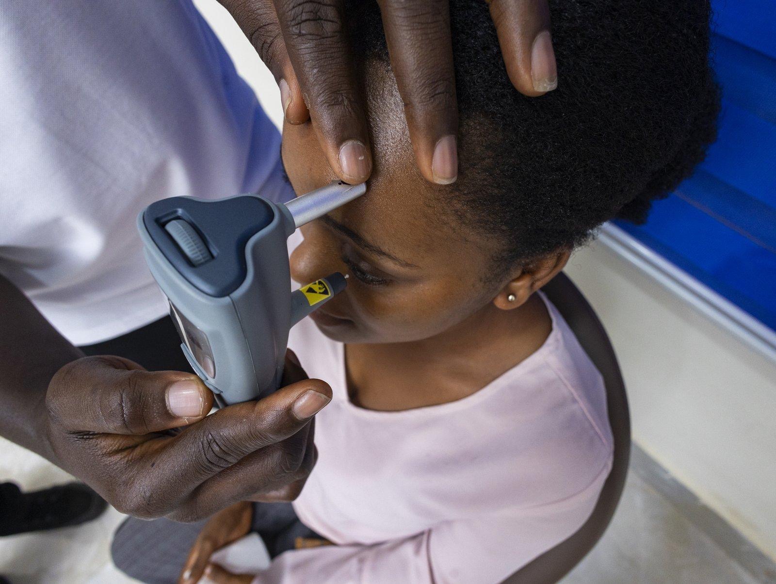
What it does: The familiar “puff of air” test that measures intraocular pressure (IOP) without touching the eye.
What it catches: Elevated eye pressure is the single biggest modifiable risk factor for glaucoma. This is usually the fastest test in any comprehensive eye exam and is repeated at almost every visit for patients being monitored for or treated for glaucoma.
Schirmer’s Test (1 and 2)
What it does: Small strips of filter paper are placed at the inner corner of each lower eyelid for five minutes, measuring how much tear film your eyes naturally produce.
What it catches: Confirms and grades dry eye disease, distinguishing between reduced tear production and other causes of dry eye symptoms, an important distinction because the treatment differs depending on the cause.
Keratometry, Amsler Grid, Colour Vision, and Stereopsis Testing
Rounding out our diagnostic suite are several more targeted tests:
• Keratometry measures the curvature of your cornea, essential before cataract surgery, contact lens fitting, or evaluating astigmatism.
• Amsler grid is a simple grid pattern used to detect distortion in central vision, often associated with macular conditions.
• Colour vision testing identifies colour vision deficiencies, which carry real-world implications, commercial drivers, for example, are required to demonstrate adequate colour vision under DVLA regulations.
• Stereopsis testing measures depth perception, particularly relevant in evaluating children for strabismus or amblyopia.
How These Tests Work Together
No single test tells the whole story.
A typical comprehensive workup for, say, a 55-year-old with a family history of glaucoma might combine tonometry (checking pressure), OCT (checking nerve fibre thickness), gonioscopy (checking drainage angle anatomy), and visual fields (confirming whether any functional vision loss has occurred).
Each test answers a different question; together, they build a complete clinical picture.
This is also why a basic vision check at an optical shop is not the same as a comprehensive eye examination at a specialist hospital. Glasses-only testing answers “can you see clearly,” while diagnostic imaging answers “is your eye healthy.” Both matter, but only one catches disease before symptoms appear.
Where to Get These Tests Done
Our full diagnostic suite is available at our Mataheko headquarters, with core imaging — OCT, fundus photography, tonometry, B-scan, and visual fields, also available at our Osu, Tema, and Kumasi branches. Some specialised tests may require referral to Mataheko, depending on the branch.
If your ophthalmologist has recommended a specific scan, or if you simply want a comprehensive baseline assessment, book an appointment and ask what’s included in a full diagnostic workup for your age and risk profile.
St. Thomas Eye Hospital’s diagnostic suite includes OCT, FFA, B-scan ultrasonography, visual fields testing, gonioscopy, fundus photography, non-contact tonometry, Schirmer’s testing, and more, available across our branches in Mataheko, Osu, Tema, and Kumasi. View all our services →
Related Reading
• 7 Laser Eye Treatments Available in Ghana
• If You Have Diabetes in Ghana, Your Eyes Are Already at Risk
• 5 Things Most Ghanaians Get Wrong About Glaucoma
• 6 Reasons Ghanaians Go Blind — and Why None of Them Have To

Dr. Michael E. Gyasi is the Medical Director and founder of St. Thomas Eye Hospital — a multi-branch specialist facility that introduced phacoemulsification cataract surgery and micro-incisional vitrectomy surgery to Ghana. Under his leadership, the hospital has delivered over 10,000 free sight restoration surgeries and screened more than 200,000 people for eye disease across the country.
Tags :