QUICK ANSWER Botox is a brand of botulinum toxin type A, a prescription medicine that temporarily reduces the activity of selected...

Experiencing a Retinal Emergency: What Ghanaians Need to Do in the Next 24 Hours

Retinal emergencies require same-day specialist assessment.
You are going about your day — maybe driving on the motorway, working at your desk, or cooking in the kitchen — when something strange happens in one eye.
A burst of bright flashes, like lightning, in the corner of your vision. Or a sudden shower of dark specks drifting across your sight that were not there an hour ago. Or, most alarmingly, a dark shadow or curtain spreading from one side of your visual field.
These symptoms can feel surreal. They do not hurt. The other eye is fine. You might be tempted to wait and see if they go away on their own.
Do not wait. These are the warning signs of a retinal detachment — a medical emergency in which the retina (the light-sensitive tissue lining the back of the eye) separates from the tissue beneath it. Without urgent surgical treatment, a retinal detachment leads to permanent, irreversible vision loss in the affected eye.
This article explains how to distinguish emergency symptoms from harmless ones, what happens when a patient presents with a retinal detachment in Ghana, what surgical treatment involves, and what recovery looks like in the Ghanaian context.
What Is the Difference Between Seeing Floaters Normally and Seeing the Floaters That Mean an Emergency?
Floaters — the small specks, dots, or cobweb-like shapes that drift across your vision — are extremely common and usually harmless. Most adults will notice them at some point in their lives, particularly when looking at a bright surface like a white wall or the sky.
Normal floaters: A few small dots or threads that have been present for weeks, months, or years. They move when your eye moves. They are more noticeable in bright light. They come and go, or you learn to ignore them. This is usually caused by age-related changes in the vitreous — the clear gel that fills the inside of the eye.
Emergency floaters: A sudden shower of many new floaters appearing within minutes or hours — often described as a “rain of pepper” or “soot” falling through the vision. These are accompanied by one or more of the following:
• Flashes of light in the peripheral vision (the sides of your visual field), often more noticeable in the dark or when you move your eyes. As the American Academy of Ophthalmology explains, flashes occur when the vitreous gel pulls on or rubs against the retina, sending false light signals to the brain.
• A dark shadow or curtain spreading across the visual field from any direction — top, bottom, or side. This is the most alarming symptom and strongly suggests that the retina has already begun to detach.
• A sudden reduction in side (peripheral) vision in one eye.
The key distinction is sudden onset and quantity. One or two new floaters that appear gradually are rarely an emergency. A sudden burst of many floaters, with or without flashes or a shadow, requires same-day specialist assessment.
How Many Hours Do I Have to Get Treatment Before Retinal Detachment Causes Permanent Blindness?
The time pressure depends on whether the detachment has reached the macula — the central part of the retina responsible for your sharpest, most detailed vision (reading, recognising faces, driving).
If the macula is still attached (“macula-on” detachment): This is a true emergency. Surgery should ideally be performed within 24 to 72 hours to reattach the retina before the macula detaches. If the macula remains intact, the chances of preserving central vision are excellent.
If the macula has already detached (“macula-off” detachment): The urgency is still high, but the timeline extends slightly. Surgery is typically performed within days to a week. The longer the macula remains detached, the less likely it is that full central vision will be recovered — though partial restoration is often still possible.
The bottom line: Do not try to diagnose this yourself. If you are experiencing any combination of sudden new floaters, flashes, or a visual shadow, contact St. Thomas Eye Hospital immediately. We can assess whether your retina is at risk and, if necessary, proceed to surgery the same day or the following morning.

Vitreoretinal surgery requires specialist equipment and trained surgeons.
Where in Ghana Can Retinal Detachment Surgery Be Performed, and What Does It Involve?
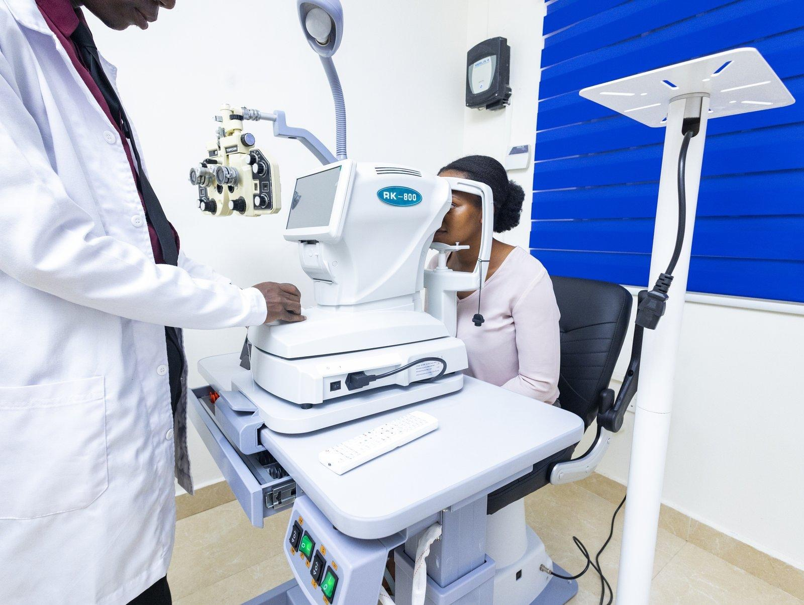
Retinal detachment surgery is a Level 3 (L3) surgical procedure that requires specialist vitreoretinal training, microsurgical instruments, and operating theatre equipment that is not available at general eye clinics or optometry practices.
In Ghana, only a small number of facilities have the capability and the trained personnel to perform these procedures.
St. Thomas Eye Hospital’s Mataheko Surgical Centre is equipped for retinal detachment surgery, including vitrectomy and combined procedures.
Surgery is performed by Dr. Michael Gyasi, the hospital’s Medical Director and fellowship-trained vitreoretinal surgeon, using a Carl Zeiss surgical microscope.
There are two primary surgical approaches to repairing a retinal detachment:
Scleral Buckling
A silicone band or sponge is sutured to the outside of the eye (the sclera), gently indenting the eye wall so that it pushes toward the detached retina. This reduces the pulling force on the retina and allows it to reattach.
Scleral buckling is often used for simpler, more peripheral detachments and can be combined with cryotherapy (freezing treatment) to seal retinal tears.
Vitrectomy
This is a microsurgical procedure in which the vitreous gel inside the eye is removed and replaced with a gas bubble or silicone oil. The gas bubble presses the retina back into place while laser or cryotherapy seals the tear.
Vitrectomy is the preferred approach for more complex detachments, detachments involving the macula, or cases where the vitreous gel has pulled scar tissue onto the retinal surface.
At St. Thomas Eye Hospital, vitrectomy is listed among our L3 surgical procedures, alongside phacoemulsification (laser cataract surgery) and glaucoma surgery. In some cases, cataract surgery and vitrectomy are combined in a single procedure known as phaco-vitrectomy.
What Is Vitrectomy and What Does Recovery Look Like in the Ghanaian Home Environment?
After vitrectomy with a gas bubble, the most important part of recovery is face-down positioning. Your surgeon will ask you to keep your head in a specific position — usually face-down or tilted to one side — for a prescribed number of hours per day, sometimes for one to two weeks.
This is not optional: the gas bubble needs to press against the retinal tear to hold the retina in place while it heals.
In the Ghanaian home environment, this can be challenging. Many of our patients sleep on mats in family compounds, share rooms with relatives, or do not have adjustable beds or specialised pillows face-down. Here is what we advise:
For sleeping: Stack several pillows on a table and lean forward, resting your forehead on the pillows. Alternatively, place a pillow on the edge of your bed and lie face-down with your face off the edge of the mattress. A folded towel with a gap for breathing works well.
For sitting: Sit in a chair with a pillow on your lap and lean forward, resting your head on the pillow. This is a comfortable position for listening to the radio, talking with family, or resting during the day.
For daily tasks: You will not be able to drive, cook over an open flame, or do heavy lifting for several weeks. Arrange for a family member or friend to help with errands and meals during the first two weeks of recovery.
Important: Do not fly while a gas bubble is in your eye. The change in air pressure at altitude causes the gas to expand, which can dangerously increase the pressure inside your eye. Your surgeon will tell you when the gas has absorbed enough for air travel to be safe (typically six to eight weeks).
After Retinal Detachment Surgery, When Can I Go Back to Work, and Are There Things I Must Not Do?
Recovery timelines vary depending on the type of surgery and whether a gas bubble or silicone oil was used:
First week: Rest at home. Attend all post-operative appointments. Use prescribed eye drops exactly as directed. Maintain face-down positioning if instructed.
Weeks two to four: Most patients with desk-based jobs can return to work after two weeks, provided they can maintain their positioning schedule and avoid heavy lifting. Patients whose work involves physical labour, dust exposure, or outdoor heat should discuss a return timeline with their surgeon.
Months one to three: Vision gradually improves as the gas bubble shrinks and is replaced by the eye’s own fluid. If silicone oil was used, a second procedure to remove the oil may be scheduled after three to six months.
Things you must avoid during recovery:
• Heavy lifting, straining, or bending at the waist
• Flying or travelling to high altitudes (if a gas bubble is present)
• Swimming or exposing the eye to unclean water
• Rubbing the operated eye
• Vigorous exercise for at least four to six weeks
What to Do Right Now
If you are reading this because you are currently experiencing symptoms, stop reading and call us now. St. Thomas Eye Hospital’s Mataheko Surgical Centre is equipped for emergency retinal assessment and same-day surgical intervention. Contact us at (+233) 54 012 5450.
If you are reading this for awareness, remember the three warning signs:
- sudden new floaters
- flashes of light
- a dark shadow or curtain across your vision.
If any of these appear, particularly in one eye, seek specialist assessment the same day. Do not wait to see if it gets better.
If you have diabetes, your retina is already at higher risk. Read our companion article: If You Have Diabetes in Ghana, Your Eyes Are Already at Risk, and ensure you are having annual dilated eye examinations.
St. Thomas Eye Hospital is one of the few facilities in Ghana equipped for vitreoretinal surgery, including vitrectomy for retinal detachment and vitreous haemorrhage. Dr. Michael Gyasi, Medical Director and vitreoretinal surgeon, received international fellowship training and performs retinal surgery at the Mataheko Surgical Centre. View all our services →
Related Reading on Our Blog
• What Causes Floaters in the Eye? A Practical Guide
• Cataract Surgery in Ghana: What Patients Ask Most
• Eye Drops, Laser, or Surgery? Understanding Your Glaucoma Treatment Options in Ghana
• 5 Things Most Ghanaians Get Wrong About Glaucoma

Dr. Michael E. Gyasi is the Medical Director and founder of St. Thomas Eye Hospital — a multi-branch specialist facility that introduced phacoemulsification cataract surgery and micro-incisional vitrectomy surgery to Ghana. Under his leadership, the hospital has delivered over 10,000 free sight restoration surgeries and screened more than 200,000 people for eye disease across the country.
Tags :