QUICK ANSWER Botox is a brand of botulinum toxin type A, a prescription medicine that temporarily reduces the activity of selected...

Retinal Detachment in Ghana: Warning Signs, Treatment, and Why Every Hour Counts
Retinal detachment is one of the few eye conditions where even a day’s delay can cost you your sight permanently.
The retina — the thin, light-sensitive tissue at the back of your eye — can separate from its supporting layer without warning. When it does, the clock starts. If the central part of the retina (the macula) detaches, the odds of recovering sharp, usable vision drop significantly with every passing day.
The challenge in Ghana? Most people experiencing the early warning signs — sudden flashes of light, a shower of new floaters, or a dark curtain creeping across their vision — don’t realise this is an emergency.
They assume it will pass. By the time they seek help, the window for a good surgical outcome may have narrowed.
This article covers what to watch for, how fast you need to act, and where in Ghana this type of surgery can actually be performed.
Normal Floaters vs. Emergency Floaters: How to Tell the Difference
Most adults will notice floaters at some point. Small specks, threads, or cobweb-like shapes that drift across your vision, especially when looking at a bright surface. These are almost always harmless clumps of protein inside the vitreous (the clear gel filling the eye).
We’ve covered this in detail in our guide: What Causes Floaters in the Eye?
Here’s the critical distinction.
Harmless floaters are few in number, have been present for weeks or longer, and don’t come with any other symptoms. They’re a nuisance, not an emergency.
Emergency floaters appear suddenly — dozens of new specks within minutes, often described as a “rain of pepper” or soot falling through your vision. They’re typically accompanied by one or more of these:
• Flashes of light in your peripheral vision, like lightning bolts. The American Academy of Ophthalmology explains that these flashes occur when the vitreous gel pulls on the retina, sending false light signals to the brain.
• A dark shadow or curtain spreading across one side of your vision. This is the single most alarming symptom. It typically means part of the retina has already detached.
• A noticeable loss of side (peripheral) vision in one eye.
The rule is simple: sudden onset, large quantity, plus flashes or a shadow = same-day specialist assessment. Do not sleep on it.
The 24-Hour Window: How Quickly Must You Act?
Time sensitivity depends on one critical factor: whether the macula is still attached.
Macula-on detachment: The retina has torn or partially separated, but the central vision area is still intact. This is a genuine emergency. Surgery should happen within 24 to 72 hours. If the macula stays attached, the odds of preserving central reading vision are excellent.
Macula-off detachment: The central retina has already separated. Surgery is still urgent — usually within a few days — but some degree of central vision loss is likely. The longer the macula stays detached, the lower the chance of full recovery.
You can’t diagnose this yourself. You can’t know whether your macula is still on or off. That’s why the response to any combination of sudden floaters, flashes, and visual shadow should be immediate: contact St. Thomas Eye Hospital and describe your symptoms. We triage retinal cases urgently.

Vitreoretinal surgery in progress at St. Thomas Eye Hospital using the Carl Zeiss S88 surgical microscope.
Where in Ghana Can Retinal Detachment Surgery Be Performed?
Retinal detachment repair is a Level 3 (L3) surgical procedure. It requires a fellowship-trained vitreoretinal surgeon, microsurgical instruments, and specialised operating theatre equipment. General eye clinics, optical shops, and most district hospitals are not equipped for this.
St. Thomas Eye Hospital’s Mataheko Surgical Centre performs retinal detachment surgery, including vitrectomy and combined procedures. Our full list of L1, L2, and L3 surgical services is available online.
Surgery is performed by specialist vitreoretinal surgeons, using a Carl Zeiss S88 surgical microscope: the same platform used at leading retinal surgery centres internationally.
There are two primary surgical approaches:
Scleral Buckling
A silicone band is sutured to the outside wall of the eye, gently indenting it to push the wall toward the detached retina. Combined with cryotherapy (freezing) to seal the tear, this approach works well for simpler, peripheral detachments. It’s been used reliably for decades.
Vitrectomy
The vitreous gel is surgically removed from inside the eye and replaced with a gas bubble or silicone oil that presses the retina back into position. Laser or cryotherapy seals the retinal tear. Vitrectomy is the standard approach for complex detachments, cases involving the macula, or situations where scar tissue has formed on the retinal surface.
In cases where cataract and retinal detachment coexist, a combined phaco-vitrectomy can address both in a single operation. This is listed among our L3 surgical procedures.
Recovery After Vitrectomy: What It Actually Looks Like in Ghana
Let’s be direct about this: recovery from vitrectomy is demanding, and it’s harder in Ghana than in countries where patients go home to adjustable hospital beds and specialised face-down cushions.
If a gas bubble was used, you’ll need to maintain face-down positioning for a prescribed number of hours each day, sometimes for one to two weeks. The gas bubble needs gravity to press it against the retinal tear. This is non-negotiable: skipping positioning risks the retina re-detaching.

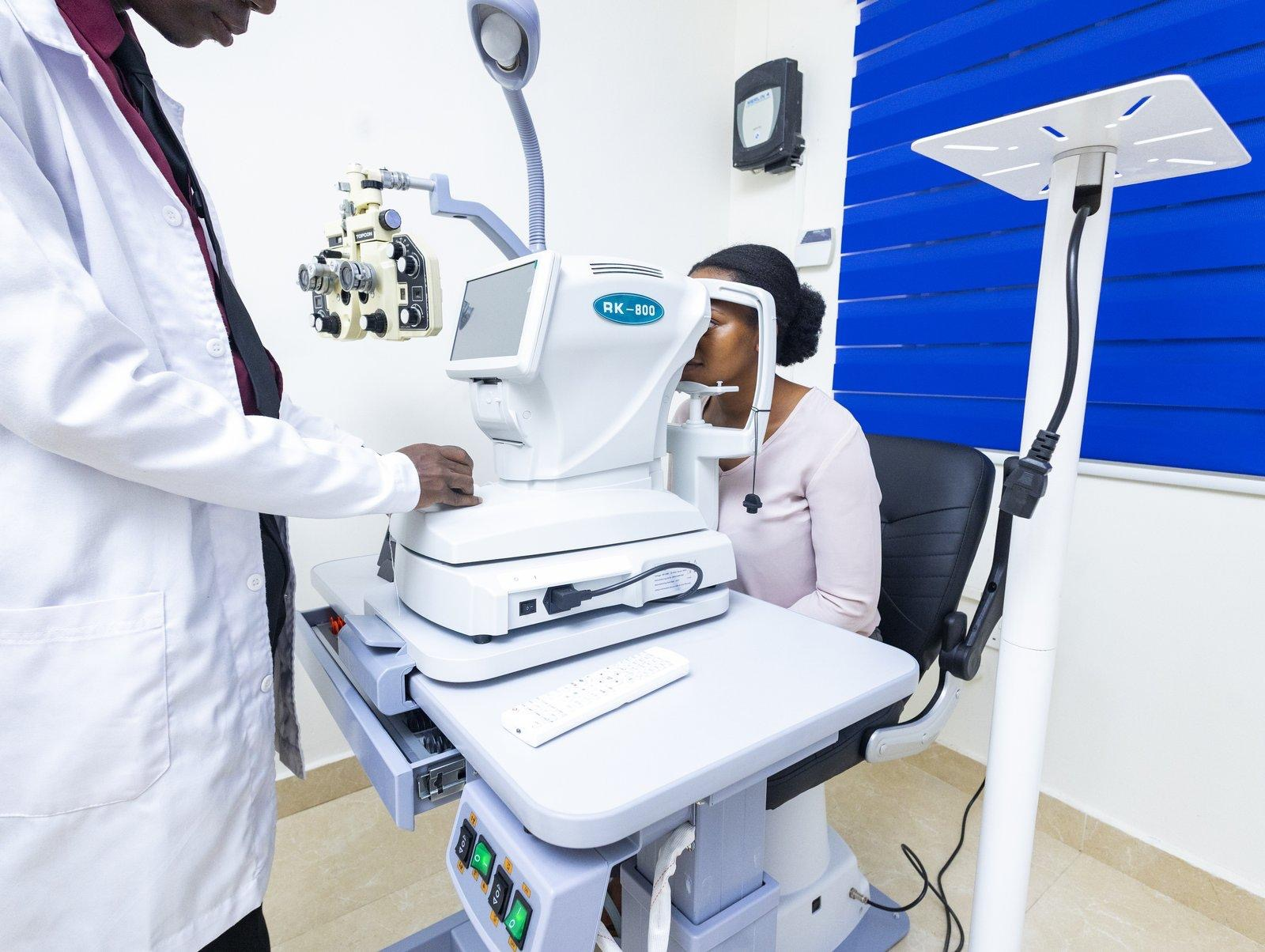
A fully equipped consultation suite at St. Thomas Eye Hospital.
Here’s how our patients make it work:
Sleeping: Stack pillows on a table and lean forward, resting your forehead on them. Or lie face-down on the bed with your face at the mattress edge, using a rolled towel with a breathing gap.
Purpose-built face-down supports exist, but most Ghanaian patients manage effectively with pillows and improvisation.
Sitting during the day: Place a pillow on your lap and lean forward. This is comfortable enough for listening to the radio, conversations, and resting for hours at a time.
Household tasks: You won’t be able to drive, cook over an open flame, or do heavy lifting for several weeks. You’ll need a family member or friend to assist, particularly in the first two weeks.
Do not fly while a gas bubble is in your eye. Cabin pressure at altitude causes the gas to expand, which can spike intraocular pressure to dangerous levels. Your surgeon will advise when the bubble has absorbed enough for air travel — typically six to eight weeks.
Return to Work and Restrictions After Surgery
Week 1: Rest at home. Attend post-operative appointments. Use prescribed drops on schedule. Maintain positioning.
Weeks 2–4: Most desk-based workers can return by week two or three, provided they can maintain positioning and avoid physical strain. Jobs involving dust, heat, or manual labour need a longer timeline — discuss with your surgeon.
Months 1–3: Vision improves gradually as the gas bubble shrinks. If silicone oil was used, a second procedure to remove the oil is typically scheduled at three to six months.
Hard restrictions during recovery: no heavy lifting or straining; no rubbing the operated eye; no swimming or dirty water exposure; no vigorous exercise for four to six weeks; no air travel until cleared.
What to Do Right Now
If you’re reading this because you have symptoms: stop reading. Call (+233) 54 012 5450 now. St. Thomas Eye Hospital’s Mataheko Surgical Centre can assess you today and operate the same day or the next morning if needed.
If you’re reading for awareness: memorise the three red flags:
- sudden new floaters
- flashes of light
- a dark curtain across your vision.
Any one of these, especially in a single eye, warrants same-day assessment.
If you have diabetes, your retina is at elevated risk. Our companion article explains why: If You Have Diabetes in Ghana, Your Eyes Are Already at Risk.
St. Thomas Eye Hospital is one of the few facilities in Ghana equipped for vitreoretinal surgery, including vitrectomy, scleral buckling, and combined phaco-vitrectomy. View all our services →
Related Reading
• What Causes Floaters in the Eye? A Practical Guide
• Cataract Surgery in Ghana: What Patients Ask Most
• Eye Drops, Laser, or Surgery? Your Glaucoma Treatment Options in Ghana
• 5 Things Most Ghanaians Get Wrong About Glaucoma

Dr. Michael E. Gyasi is the Medical Director and founder of St. Thomas Eye Hospital — a multi-branch specialist facility that introduced phacoemulsification cataract surgery and micro-incisional vitrectomy surgery to Ghana. Under his leadership, the hospital has delivered over 10,000 free sight restoration surgeries and screened more than 200,000 people for eye disease across the country.
Tags :